Chapter 5
Effects of sustained acceleration on the morphological properties of otoconia in hamsters
Published in: Acta Otolaryngologica (Stockholm). 115:227-230, 1995
Abstract
We investigated the effect of prolonged
hypergravity on the otoconial layer of the maculae utricles and the maculae
saccules in hamsters. The animals were placed in a centrifuge under conditions
of 2,5 G and stayed there for six months. We then determined the calcium contents
of the otoconia with energy dispersive X-ray element analysis and recorded the
size, shape and distribution of the otoconia. Scanning electron microscopy was
used to make photos to determine the effects of hypergravity on the shape and
size of the otoconia and the distribution of zones that contain smaller and
larger otoconia.
No differences were found in the calcium content, shape, size and distribution
of otoconia between centrifuged hamsters and controls. Our findings indicate
that structural adaptation to hypergravity does not take place at the otoconial
level, at least not in animals which were subjected to hypergravity after the
vestibular system was fully matured.
Keywords: hypergravity, scanning electron microscopy, EDAX element analysis, vestibulum, utricle, sacculus, otoconia, hamsters.
Introduction
The vestibulum contains the macula sacculus
and the macula utricle. Both maculae are designed to detect linear accelerations
such as gravity. The superficial layer of both maculae is capped with otoconia,
which are crystals containing calcium carbonate. Otoconial are formed during
the second half of gestation. At birth, the otolith organs seem fully matured
(Sánchez-Fernández et al. 1984; Kawamata and Igarashi, 1993; Kido
et al. 1993). Ross et al (1987) suggested that the otoconia represent seismic
masses that are spring loaded through the underlying otolith membrane, to the
cilia and some of the longer stereocilia.
The otoconia, as weight-lending structures, could be subjected to changes when
exposed to prolonged increased gravity (hypergravity). The extra G-load may
cause an increase in the weight of the otoconia. If structural adaptation occurs,
the deposition of calcium carbonate in the otoconia may be reduced during hypergravity.
This would result in a lower weight of the otoconial mass. Howland and Ballarino
(1981) found that in embryos developed in hypergravity (1.8 - 2.1 G) for more
than 17 days, the weight of the utricular macula was decreased when compared
to normal embryos raised in a 1 G environment. They suggested that the decrease
was caused by a reduction in the calcium content of the otoconia. Other investigators
have suggested that long-term exposure to altered gravity may also alter the
shape, size or distribution of the otoconia on the macular disk (Lim et al.
1974; Vinnikov et al. 1979; Ross et al. 1985; Ross, 1987; Krasnov, 1991).
The aim of this study was to investigate the effect of prolonged hypergravity
on the otoconial layer of the utricular and saccular maculae in hamsters. We
also determined the calcium contents of the otoconia and their size, shape and
distribution.
Methods
Centrifuge
The animal centrifuge consisted of a centrally- placed 3.5. kW DC motor drive and 2 horizontally mounted arms of 1.15 meter long. Each arm was connected to an aerated and darkened free-swinging gondola (length 110 cm, width 45 cm, height 80 cm). During centrifugation, the Z-vector was constantly directed to the floor of the gondola. A rotation speed of 34,3 RPM produced a 2.5 G-value was at the floor of the gondola. A video camera in the gondola made it possible to observe the animals' behaviour.
Animals
Golden hamsters (Mesocricetus auratus) were obtained from Harlan (Zeist, the Netherlands). Ten hamsters (21 days old) were placed in acrylate boxes, which were loaded into the centrifuge gondola. During the experiment the hamsters lived under conditions of 2,5 G (hypergravity or HG hamsters). Ten control hamsters lived in similar housing conditions in normal gravity in the same experimental room. Food and water was available ad libitum. For six months, centrifugation of the hamsters was continuous except for one half hour per day for animal care and testing of the perceptive-motor skills, such as maintenance of equilibrium and orientation. The results of the behaviour studies are presented elsewhere.
Histology
After 6 months, the hamsters were killed
and the temporal bones of HG hamsters and control animals were dissected. The
maculae utricle and saccule, were fixed in 2.5% gluteraldehyde + 0.5% paraformaldehyde
in phosphate buffer solution (0,1 M, pH 7.4). The specimen were rinsed in distilled
water and air-dried, and were then prepared for calcium content analysis and
scanning electron microscopy (SEM, ISI 40).
To determine the calcium contents of the otoconia, the specimen were sputter-coated
with carbon and subjected to energy dispersive X-ray (EDAX) elemental analysis.
For SEM observation, the specimen were mounted on aluminium stubs and coated
with gold. Photos were made to determine the effects of hypergravity on the
size of the otoconia and the distribution of otoconia in three different zones
containing small medium and larger otoconia. The distribution of different sizes
was determined with the help of a MOP-Videoplan XY digitalizing tablet (Kontron,
Munich, Germany) with software.
Results
Elemental analysis.
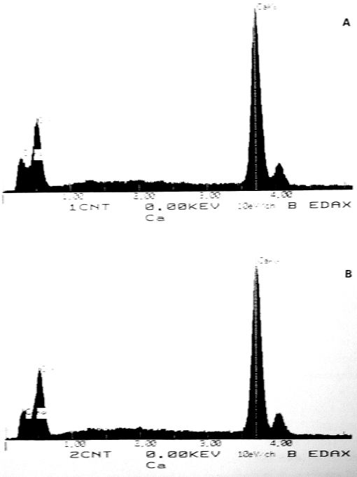
For EDAX element analysis utricular and saccular patches were used from three HG hamsters and three control hamsters; in all 12 utricles and 12 saccules were analyzed. Calcium content was the same in otoconia of both the utricle and the sacculus. Calcium peaks were detected for both groups with no differences in the height of the peaks. (Fig. 1). We found no differences in calcium content between large otoconia and small otoconia. Element analysis revealed no differences between otoconia from HG hamsters and otoconia from control hamsters. Calcium peaks were detected in both groups, and there were no difference in the height of the peaks.
Fig. 1. Calcium content of otoconia of the sacculus of (A) the control hamster; (B) the hypergravity hamster. Highest peak represents calcium.
Size and distribution of the otoconia.
Micrographs of the otoconial layer of the
utricle and saccule of HG hamsters and control hamsters showed that utricular
otoconia and saccular otoconia were similar in size in HG hamsters and control
animal. Furthermore, small, medium-sized and large otoconia were found in the
utricle of both HG hamsters and controls. The small and medium-sized otoconia
were located in the center of the otoconial patch whereas large otoconia were
found at the periphery of the patch (Fig. 2). Anomalies in the shape of the
otoconia, such as two or more otoconia fused together, were found in the utricle
as well as in the saccule (Fig. 3) of both groups.
With the help of an XY digitalizing tablet, the surfaces of zones of the utricle
covered with small, medium-sized and large otoconia were measured in a double
blind fashion by three observers (6 intact patches from control hamsters and
13 patches from HG hamsters). The percentage surface of each zone in proportion
to the total surface of the otoconial layer was calculated (one-way analysis
of variance), and the results of the three observators were compared. Although
considerable variation was found in the distribution of the three sizes of otoconia
from one animal to another, the distribution of otoconia of the macula utricle
(figure 4) was almost identical in HG and control hamsters (small: F(1,55)=.25,
p=0.62; medium: F(1,55) <0.01, p= 0.98; large: F(1,55)= 0.07, p=0.79).
Discussion
In this experiment, the calcium content
of the utricular otoconia was the same as in the saccular otoconia, which is
in agreement with the results of Campos et al (1992). We found no differences
in calcium content between smaller and larger otoconia, in contrast with findings
of Campos et al (1984). This discrepancy may be due to the difference in the
ages of the animals studied: Campos and colleagues studied newborn rats whereas
we used adult hamsters.
Hypergravity did not affect the calcium content of otoconia in HG hamsters in
comparison to controls. Furthermore, in both groups the calcium content was
the same in utricular and saccular otoconia. Although Howland and Ballarino
(1981) used chick embryos whereas we used used adult hamsters, the reduction
in the mass of the utricular maculae found by the former was probably not caused
by a reduction in calcium content of the otoconia but rather by a decrease in
the mass of underlying tissue such as the neuroepithelia.
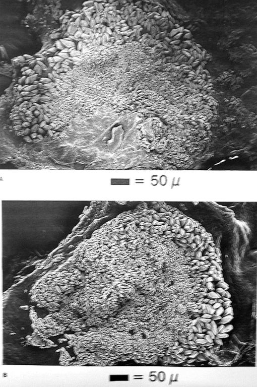
Fig. 2. Otoconial layer of the utricle of (A) the control hamster. The otoconial patch is partly covered with membrane; (B) the hypergravity hamster.
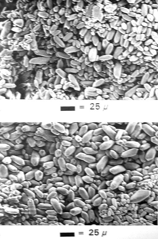
Fig. 3. Otoconia of the sacculus of (A) the control hamster; (B) the hypergravity hamster.
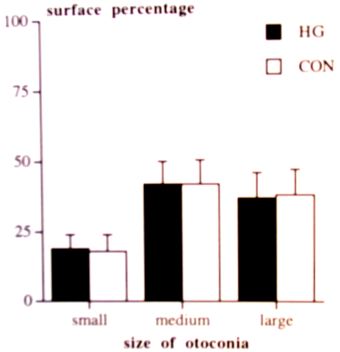
Fig. 4. Percentage surface distribution of small, medium and large otoconia of the utricle of control hamsters and hypergravity hamsters. Shown are means and standard deviations.
In this study, the shape of the utricular
and saccular otoconia of the HG hamsters was not changed. Moreover, other studies
of the effect of prolonged hypergravity did not report any changes in the shape
of the utricular or saccular otoconia (Krasnov, 1991). During microgravity,
alterations in the shape of otoconia were found; for example, Vinnikov et al.
(1979) reported that otoconia became oval and rounded in shape in rats subjected
to weightlessness for 20 days. They also found changes in the distribution of
light (organic) and dark (inorganic) substances of the otoconia. Ross et al
(1987) found accumulations of very small otoconia in the peripheral zone of
the utricular otoconial patch and a smoothing out of saccular otoconial body
surfaces in mature rats subjected to weightlessness for 7 days. A comparison
of the results of the microgravity experiments with the results of hypergravity
experiments suggest that the effects of hypergravity are not simply the opposite
of those observed when the G force is absent, as during microgravity. However,
the microgravity experiments lasted shorter than the hypergravity experiments
and the methods used to measure the otoconia are not described.
No differences in the size of the utricular or saccular otoconia were observed
between maculae of HG hamsters and control animals. Furthermore, no changes
in the distribution of the otoconia on the utricular patch were observed in
HG hamsters in comparison with controls. The percentage surface containing small,
medium or large otoconia was the same in both groups. In contrast with our findings,
Krasnov (1991) found that the formation of large otoconia was inhibited in the
anterior third part of the periphery of the otolith membrane of rats developed
pre- and postnatally under conditions of 2 G. Hara (1993) found giant otoconia
in the peripheral zone of the utricular patch in chick embryos exposed to hypergravity
(2G) during the embryonic period, which is the opposite effect of that found
by Krasnov (1991). It should be noted that alterations in the distribution of
the otoconia, like those described by Krasnov (1991), were observed only in
a small part of the otoconial layer. Therefore, the Krasnov's view that prolonged
hypergravity inhibits the formation of large otoconia in the utricle is not
supported by the present data.
The results of this study indicate that otoconia show no structural adaptation
to hypergravity, at least not in animals subjected to hypergravity after the
otoconial genesis was complete. However, structural adaptation may still take
place within the otolith organs after maturation. Adaptation can also occur
at a different level such as the subcupular meshwork or the sensory epithelium.
Further studies are needed to determine whether structural adaptations to hypergravity
occur in the otolith organ occurs either during development or after maturation
of the vestibular system.
Acknowledgements
We thank BLJ Willekens of the Department of Morphology (Ophthalmic Research) for the EDAX element analysis. The Netherlands Organization for Scientific Research (NWO) is gratefully acknowledged for funding this project. This research was conducted while HNPM Sondag was supported by a grant of the Foundation for Behavioural and Educational Sciences (SGW) of this organization (575-62-049), awarded to Prof. Dr. WJ Oosterveld.